Ultrasound General
Ultrasounds are safe and efficient diagnostic tools using sound waves to create images of certain parts of your body. We offer a broad range of the ultrasound evaluations including obstetrical, abdominal, and male and female pelvic studies.
We also perform many other studies including transrectal (prostate), breast, scrotum and thyroid studies. We specialize in musculoskeletal ultrasound, such as assessment of the shoulder, knee, hip, ankle, foot, wrist and other joints. We can assess soft tissue injuries, including occupational and sport trauma related problems.
Ultrasound is safe, noninvasive, and does not use ionizing radiation.
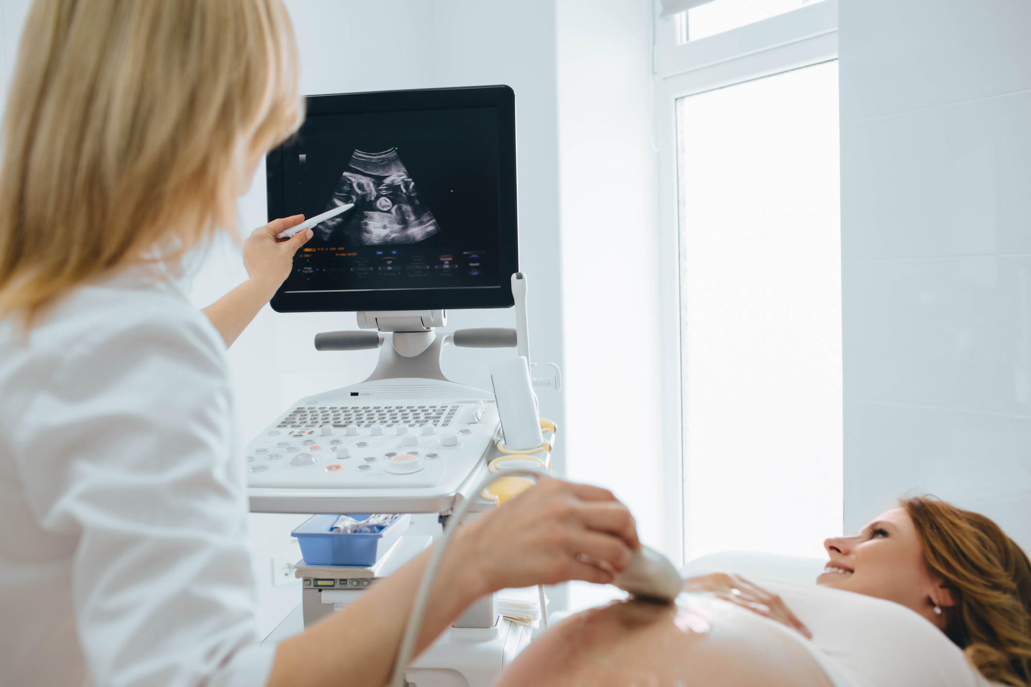
Pregnancy 1st Trimester Scans: A dating scan is performed around week 8 of pregnancy but can be done as early as 5 weeks. It is used to confirm due dates, assess the viability of the pregnancy, check the number of embryos, provide maternal reassurance, and to rule out ectopic pregnancy (fetus developing outside of the womb). Nuchal Translucency (NT) Scan Following the dating scan, a Nuchal Translucency (NT) scan is usually booked in around week 12 of pregnancy but can generally be done between 11 weeks to 14 weeks. The NT scan is a screening test for early detection of Down syndrome. A blood test is also done in conjunction with the ultrasound scan which is arranged through the patient’s Lead Maternity Carer (LMC). Early fetal anatomy can also be assessed during this scan. 2nd Trimester scans: At 18 to 20 weeks of pregnancy, an anatomy scan is performed to rule out abnormalities that can be visualised with ultrasound imaging such as cleft lip, spina bifida, heart defects, and many other abnormalities. It is also used to check that the fetus size is within normal limits and record the location of the placenta. The gender of the fetus can often be established during this scan. 3rd Trimester Scans: Usually conducted from week 28 of pregnancy, the growth scan is used to check the baby’s growth by measuring its head, abdomen, and thigh bone. It is also used to assess the amount of amniotic fluid surrounding the baby and record the position of the placenta. |
MusculoskeletalShoulder Scan An ultrasound scan of the shoulder is used to check for muscle tears and fluid associated with the rotator cuff, swelling and subluxation of the acromioclavicular (AC) joint, and the long head of the biceps tendon (LHB) can be assessed. Ankle Scan An ultrasound scan of the ankle is used to assess tendons and ligaments for damage and also the ankle joint for fluid. Ruptures, tears, and tendinopathy of the Achilles tendon and Plantar Fascia can also be well assessed with ultrasound imaging. Knee Scan An ultrasound scan of the knee is used to assess the Medial Collateral Ligament (MCL), Lateral Collateral Ligament (LCL), quadriceps and patella tendon for strains and tears. The popliteal fossa is also checked for Baker’s cysts and other abnormalities. Elbow Scan An ultrasound scan of the elbow is used to rule out lateral epicondylitis (tennis elbow) and medial epicondylitis (golfer’s elbow) and assess injury to the lower parts of the biceps and triceps tendon. Wrist Scan An ultrasound scan of the wrist is used to check for swelling of the tendons and ligaments in the wrist, assess the nerve pathology. De Quervain’s abnormality is a common condition of the wrist that is diagnosed with ultrasound imaging. Calf muscle Scan Tears in the calf muscle region can be well visualised with ultrasound. Lumps and bumps An ultrasound scan is also used on a variety of palpable lumps and bumps to assess their nature. |
General UltrasoundAbdominal Ultrasound An ultrasound scan is used to assess the pancreas, aorta, liver, gallbladder, bile ducts, kidneys and spleen. It is also used to check for fluid in the abdomen (ascites). Gallstones, liver and kidney cysts, aortic aneurysms and other abnormalities can be assessed with ultrasound. Pelvic Ultrasound (female) An ultrasound scan is used to view the uterus (womb) and ovaries for things such as ovarian cysts, endometrial disorders, fibroids, Intrauterine Contraceptive Device (IUCD) location, or other disorders for pelvic pain and other symptoms. Renal Ultrasound An ultrasound scan is used to assess the urinary bladder and kidneys to check for stones, blockages, lesions and other abnormalities. Prostate Ultrasound Ultrasound of the prostate uses sound waves to produce pictures of a man’s prostate gland and to help diagnose symptoms such as difficulty urinating or an elevated blood test result. It’s also used to investigate a nodule found during a rectal exam, detect abnormalities, and determine whether the gland is enlarged. |
Small Parts UltrasoundThyroid Scan An ultrasound scan of the thyroid is used to check for cysts, nodules, and abnormalities and to assess the size and offer follow-up assessment. Breast Scan An ultrasound scan of the breast is used to assess a palpable lump or to monitor the size of previously seen lesions. Scrotal Scan An ultrasound scan of the scrotum is used to assess a palpable lump, testicular pain, epididymal lumps or abnormalities. It is also used to assess the scrotal size and to rule out testicular torsion, hydrocele and varicocele. |